Otago gains state-of-the-art health research microscope

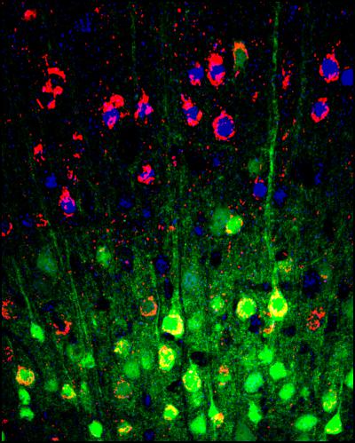
Image shows the organisation of brain cells (neurons) in a part of the brain responsible for movement, the motor cortex; the green neurons provide the electrical signals that drive movement of the limbs.
Monday 14 April 2014
Otago gains state-of-the-art health research microscope
University of Otago medical scientists will now be able to peer into the cells of the living brain for the first time to study the onset and progression of such neurological diseases as Parkinson’s and Alzheimer’s.
This is thanks to a newly acquired Multiphoton Microscope which applies a powerful but harmless infra-red light that literally sees into living organs and cells with unparalleled detail and speed. An official launch for the Microscope is being held on 15 April in the University’s Lindo-Fergusson Building.
Associate Professor Ruth Empson of the Department of Physiology and the University’s Brain Health Research Centre, says the multiphoton microscope will be used to image a wide variety of living tissues and organs including the brain, skin, lungs, gut and lymph nodes.

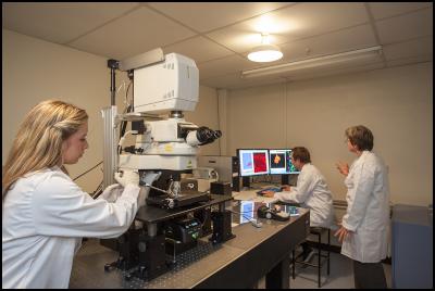
Researchers at work with the microscope
“It is very appropriate that the Department of Physiology is driving this initiative since physiology is the scientific discipline that aims to understand how living things work,” she says.
“Understanding how they work is critical for fixing them, so the knowledge developed with this new microscope will help answer important questions in human health and diseases including stroke, arrhythmia, wound healing and irritable bowel disease.”
She says that having the ability to see deep into a previously impenetrable structure like the brain, and measure its electrical activity, will revolutionise our understanding of how complex networks of brain cells use electrical impulses to communicate with one another.
“We will be able to see how these impulses change or malfunction in response to a brain trauma such as a stroke, or diseases such as Parkinson’s or Alzheimer’s. We will also be able to visualise changes in brain structure during development, across puberty, right through to old age.”
In addition to its use in brain research, the multiphoton microscope will also provide the ability to visualise activities of other organs such as the heart, allowing Otago scientists to understand how changes in heart tissue can lead to cardiac arrhythmias.
Another application will be to visualise the cells through the lining of the gut for the treatment of inflammatory bowel disease.
The first of its kind in New Zealand, the $1 million multiphoton microscope is one of only a handful throughout the world. It was acquired with the assistance of NZ Lottery Health funding as well as a variety of internal University of Otago sources.


 Science Media Centre: Cyclone Gabrielle's Impacts On NZ's Ecosystems - Expert Reaction
 RNZ: Parts Of Power System Could Be Out For 36 Hours In Event Of Extreme Solar Storm
 NZAS: New Zealand Association Of Scientists Awards Celebrate The Achievements Of Scientists And Our Science System
 Stats NZ: Retail Spending Flat In The September 2024 Quarter
 Antarctica New Zealand: International Team Launch Second Attempt To Drill Deep For Antarctic Climate Clues
 Vegetables New Zealand: Asparagus Season In Full Flight: Get It While You Still Can


